
1. 대회 데이터

각 독립 사례에는 5자리 숫자로 식별되는 전용 폴더가 있습니다. 이러한 각 "케이스" 폴더에는 DICOM 형식의 각 구조적 다중 파라미터 MRI(mpMRI) 스캔에 해당하는 4개의 하위 폴더가 있습니다.
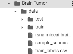

식별 넘버 5자리 숫자가 train 안의 폴더 이름들. 저런걸 말하나 보네요.

각 식별 번호 폴더 안에는 다음과 같은 4개 폴더가 있습니다.
그 안에도 이미지로 꽉 차 있어요.
- Fluid Attenuated Inversion Recovery (FLAIR)
- T1-weighted pre-contrast (T1w)
- T1-weighted post-contrast (T1Gd)
- T2-weighted (T2)
T1Gd 가 바로 T1wCE를 말하는 것 같습니다.
위 용어에 대한 설명은 다음 게시글에서 따로 정리하도록 하겠습니다.
2. RSNA 공식 홈페이지에서 설명된 데이터
| Imaging Modality and Contrast | MRI ● Pre- and post-contrast |
| Annotation Pattern | ● 3D VOI(s) |
| Annotation methodology and structure | Method of annotation ● Manual Annotation output ● Spreadsheet (text) ● Bounding boxes ● DICOM (e.g., SR DICOM) ● JSON ● Proprietary ● NIfTI Annotation software ● Not specified Storage, Portability, Interoperability ● RSNA/Synapse storage (Task 1) and Kaggle (Task 2) |
| Structure nomenclature and standards | ● None (Organizer-specified) |
| Data use agreement/licensing | ● Open Licensing ● Non-commercial purpose ● Registration (e.g., the intent of use) ● References to dataset ● Versioning |
| Imaging file and structure set format | ● DICOM - metadata/tags (based on individual task) ○ Proprietary tags ● NIfTI ● PNG/JPEG/TIFF ● Raw data ● Other |
| Slice thickness (in mm) | Variable |
| Characteristics | Resolution (해상도) ● Original (원본) ● Downsampled (다운샘플링) Pre-processing (전처리) ● Standard Normalization (표준 정규화) ● Histogram Normalization (히스토그램 정규화) ● Skull stripping (두개골 박리) Burned-in PHI ● Removed |
| Other scanner and acquisition parameters, e.g. MRI field strength |
Available but incomplete (missing 0008,0070 Manufacturer) |
| Timing (in case of serial imaging) | ● Timepoint 1, Timepoint 2, etc. ● In relation to a relevant event ○ Pre-treatment ○ Mid-treatment ○ Post-treatment |
| Labeler demographics | ● Specialty indicated ( 특이사항 표시 ) ● Extract individual (개인정보 추출 ) ● Scope of annotation - multi-institutional (주석 범위 - 다중 기관) |
| Responsibilities quality, safety, privacy | Indicated |
| Monetization (수익 창출) | None |
* 두개골 박리 : 두개골 박리는 뇌의 이상을 감지하는 과정의 초기 단계 중 하나이다. 뇌의 MRI 이미지에서 비뇌 조직으로부터 뇌 조직을 분리하는 과정이다.

아래 링크 통해 가져왔습니다.
Brain Tumor AI Challenge (2021)
Task 2: Brain Tumor Radiogenomic Classification Participants built models that use mpMRI imaging to predict MGMT promoter methylation status, an important biomarker for treatment of brain tumors. Such radiogenomic models could improve the efficiency and ac
www.rsna.org
어려운 용어가 많네요..
틈틈히 정리를 하도록 하겠습니다.
데이터 인용 :
U.Baid, et al., “The RSNA-ASNR-MICCAI BraTS 2021 Benchmark on Brain Tumor Segmentation and Radiogenomic Classification”, arXiv:2107.02314, 2021.
'Brain Tumor Radiogenomic Classification' 카테고리의 다른 글
| [Brain Tumor] DICOM 파일이란? (1) | 2022.09.30 |
|---|---|
| [Brain Tumor] T1W, T2W, T1Gd, Flair 영상 비교 - 용어정리 2 (0) | 2022.09.29 |
| [Brain Tumor] MRI, T1 강조, T2 강조 - 용어 정리 1 (0) | 2022.09.28 |
| [Brain Tumor] 대회 설명 (0) | 2022.09.28 |
| Brain Tumor Radiogenomic Classification 프로젝트 (0) | 2022.09.28 |